ADAS 3D LV
How can ADAS 3D help you?
01.
Help in deciding the approach
02.
Understanding of LV substrate
We help you to understand the LV substrate by visualizing and quantifying its fibrotic tissue distribution.
03.
Increase the operator’s confidence
We help increase the operator’s confidence during navigation by providing a roadmap to assist in the procedure.
04.
Obtain all the benefits from advanced imaging
We help you to connect to advanced imaging radiological services to import DICOM images for MRI.
How it works
Decision making before intervention
Understand the fibrosis distribution in the myocardium from MRI and the wall thickness and surrounding anatomical structures from CT.
During the procedure
Use the ADAS 3D model as a roadmap to the catheter navigation system to accurately treat your patient.
Post-procedure revision*
After the intervention, study the electroanatomical map with ADAS 3D and compare it with the MRI information to quantify outcomes. *Not available for clinical use in the USA
Analysis available for MRI and CT images
ADAS 3D processes both MRI and CT. Magnetic Resonance provides information about the fibrotic tissue. Computed Tomography provides detailed information about myocardial wall thickness and heart anatomy.
With MRI, you can obtain:
Analysis from LV Enhancement:
- Visualization of the left ventricle in concentric layers from endo to epi
- Detection of anatomical corridors
- Identification of 3D core tissue
Heart Anatomy Extraction:
- coronary arteries, aorta, esophagus or other heart structures
With CT, you can obtain:
- Heart Anatomy Extraction such as coronary arteries, aorta, esophagus or other heart structures.
- LV Wall Thickness
The technology
Features
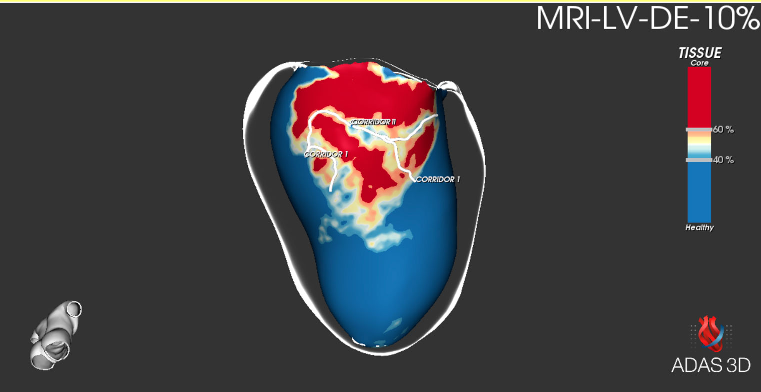
Border-zone corridors automatically detection
Automatic transmural detection of border-zone corridors and the quantitative analysis for each corridor.
Easy-to-understand ‘layers’
Visualize the Left Ventricle in concentric layers going from endo to epicardium to appreciate the scar transmurality.
Tissue characterization
Obtain a 3D Cardiac Model Segmentation with an automatic classification of tissue using selectable imaging thresholds
Export to EP navigation system
Help during the intervention by exporting structures (including anatomy, fibrosis information and corridors) to formats compatible with EP navigation systems
Technical characteristics
Windows-based
ADAS 3D software can be installed in either a laptop or desktop PC (see minimum requirements).
Connection with the PACS
Retrieve DICOM and store your cases from a centralized archive. Available in Cvi42 integrated version.
Floating licenses
Access to ADAS 3D from any computer in the same hospital with one license. Available in Cvi42 integrated version.
Study Storage Database
Storage and management of processed studies.
Try the interactive demo
LV 2D DE-MRI
Discover the interactive 2D view of the LV DE-MRI and explore the tissue characterization.
LV 3D DE-MRI
Discover the interactive 3D view of the LV DE-MRI and explore the tissue characterization.
LV-CTA
Discover the interactive 3D view of the LV-CTA and explore the tissue characterization.
Customer Portal
Find all the information related to ADAS 3D product.
Instructions For Use
Refer to the user manual to learn the features available and how to use the software.
Training tutorials
For educational purposes, we have developed video tutorials and practice cases. Use them for the initial steps on your training.
Technical support
Regarding the installation process, consult the PC requirements and the license request to start using ADAS 3D.
Image acquisition recommendations
High-quality images are necessary for the analysis of the fibrosis. Obtain ADAS 3D compatible DE-MRI and CTA sequences.
Scientific evidence
We provide you with a list of publications and articles related to ADAS 3D. Check the selection of the most relevant studies published in the last years.
Help to process a case
Our team guides you through all the steps. Have a look at the general workflow on how to obtain feedback and request help for a case.
Product media
Find some examples of real life cases using ADAS 3D.
Screenshots
Videos
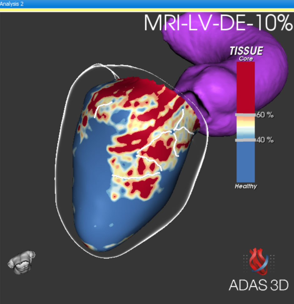
Fibrosis Analysis
ADAS 3D model with a tissue characterisation in reference to fibrosis analysis obtained from MRI. Anatomical corridors are also represented in the ventricle.
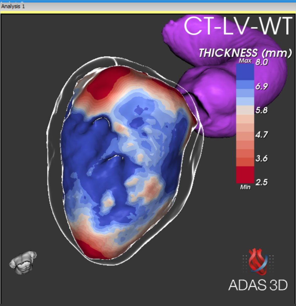
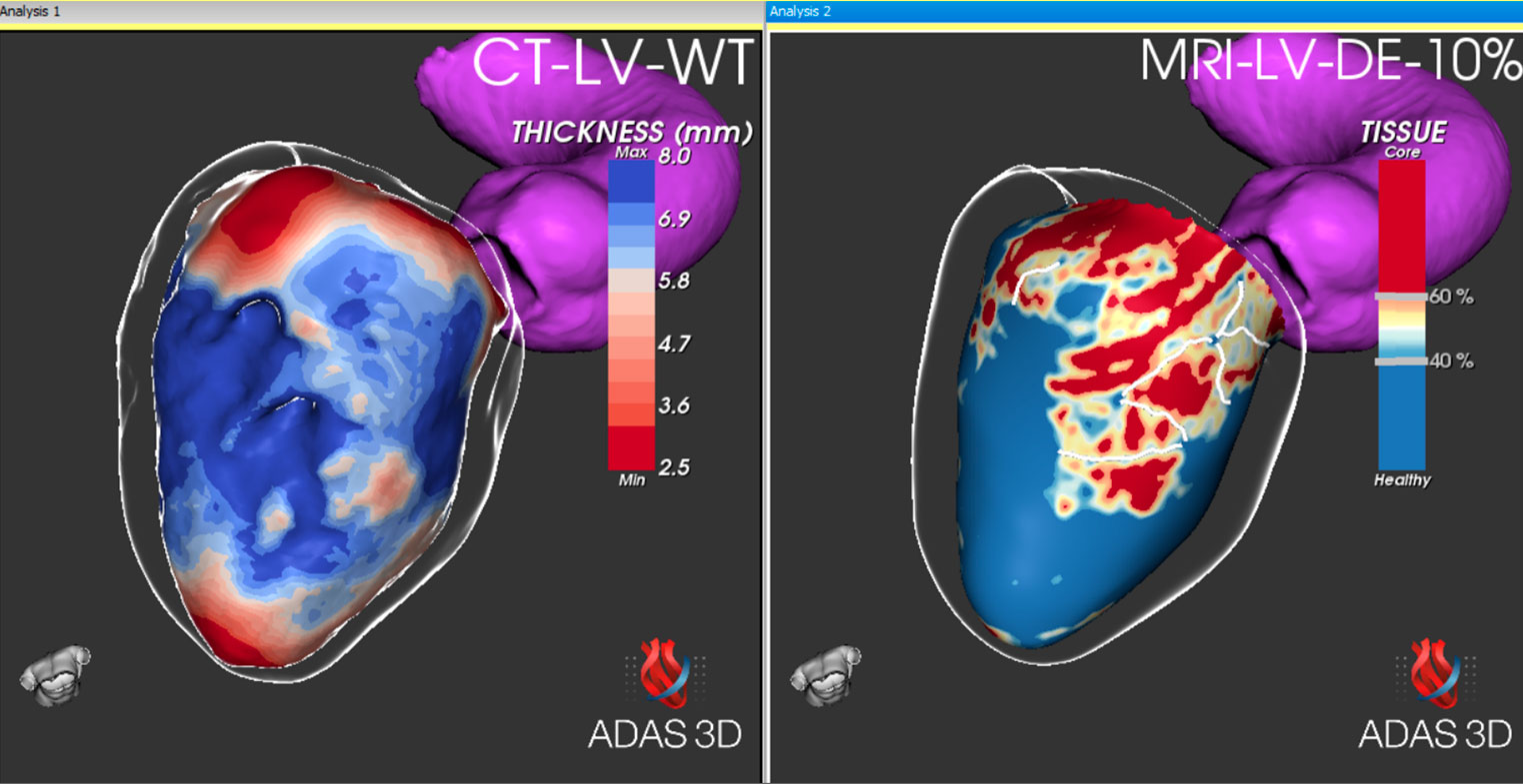
Wall thickness and Anatomy
Correlation between DICOM image and ADAS 3D model. The user can observe the Wall Thickness analysis and anatomical information obtained from CT.
Correlation between DICOM and 3D Model
DICOM Late Gadolinium Enhanced Cardiac MRI data is displayed on the right. On the left, ADAS 3D software depicts correlation with the 3D model of the left ventricle.
Concentric layers of LV
The tissue is displayed in concentric layers going from endocardium to epicardium. The user can analyse scar transmurality and the presence of corridors.
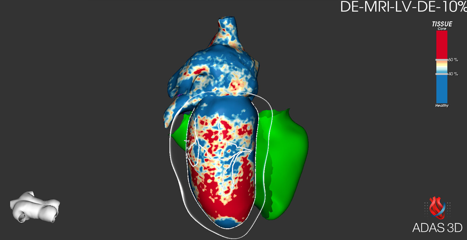
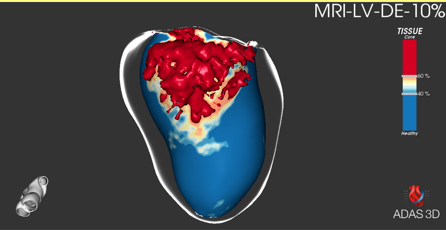
Fibrosis represented in 3D
According to tissue characterisation, fibrotic core zone is represented in 3 dimensions.
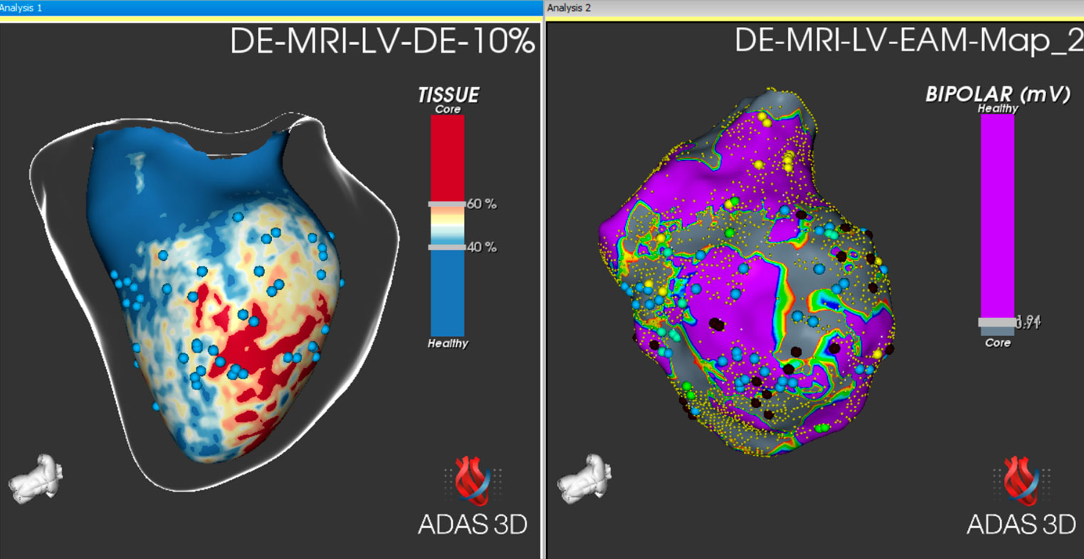
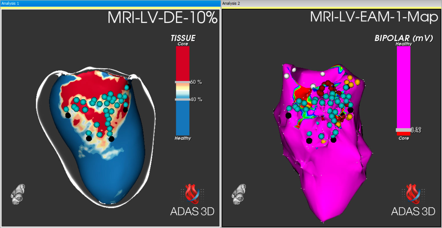
EAM revision
After the intervention, study the electroanatomical map with ADAS 3D and compare it with the MRI information to quantify outcomes. *Not available for clinical use in the USA
Testimonials
Clinically evaluated in centers worldwide
ADAS 3D LV features are clinically validated in collaboration with hospitals and universities all over the world.