We present a case from Dr. Alba Santos (Hospital Vall d’Hebron, Spain) of a patient suffering from ventricular tachycardia (VT). The magnetic resonance images (MRI) and computed tomography (CT) were analyzed with ADAS 3D software to extract detailed information about the patient.
Description of the case
We present a 50-year-old woman with a history of inferior myocardial infarction with no significant lesions in the coronary arteries and mechanical mitral valve prosthesis because of severe mitral regurgitation. A single chamber ICD was implanted after recovered sudden cardiac death ten years ago.
Recently, she received ICD therapies due to ventricular tachycardia, which has recurred despite antiarrhythmic drugs. Due to the presence of a mechanical mitral prosthesis, retrograde aortic access was used for ablation.
Analysis with ADAS 3D software
Before the procedure
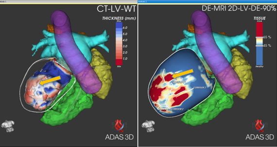
The DICOM images were imported into ADAS 3D software to be processed by Gabriel Pascual Gonzalez (nurse at Hospital Vall d’Hebron, Spain). The tissue characterization analysis of the left ventricle was performed using the DE-MRI. The DE-MRI had some artifacts caused by the device, which were excluded from analysis, luckily not affecting the area of interest.
LV wall thickness map and some anatomical structures (left atrium, device cables, calcifications, prosthetic mitral valve, the aorta, right ventricle, right atrium, and pulmonary artery) were obtained from the CT.
An interactive ADAS 3D model which displays the tissue characterization analysis from the DE-MRI can be found in:
https://www.adasonline.com/open?study_id=894911ea-ea4f-4181-8a5c-13a2548bfa64
An interactive ADAS 3D model which displays the tissue characterization analysis from the CT with the anatomical structures can be found in:
https://www.adasonline.com/open?study_id=d3ac9f5f-5060-495e-9a60-7af9e6b809e0
During the procedure:
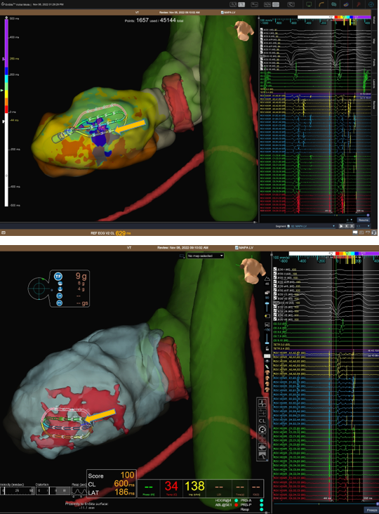
During the procedure, the analysis of ADAS 3D was imported into the Ensite X navigation system, and the electro-anatomical map was done with the HD grid catheter. In the image below, the EAM is shown with the HD grid in the Ensite X together with the prosthetic valve, device lead, calcifications, and the left atrium from the ADAS 3D analysis. The visualization of the calcifications and the prosthetic valve, helped to be more careful during the procedure near these areas.
After the procedure:
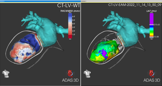
After the procedure, the EAM was imported into ADAS 3D and compare with the CT analysis. In the following image the high correlation between the CT analysis (displaying the areas of thinning and the calcifications) and the EAM can be seen. In addition, the calcifications are visualized in the area of interest.
What does the expert say?
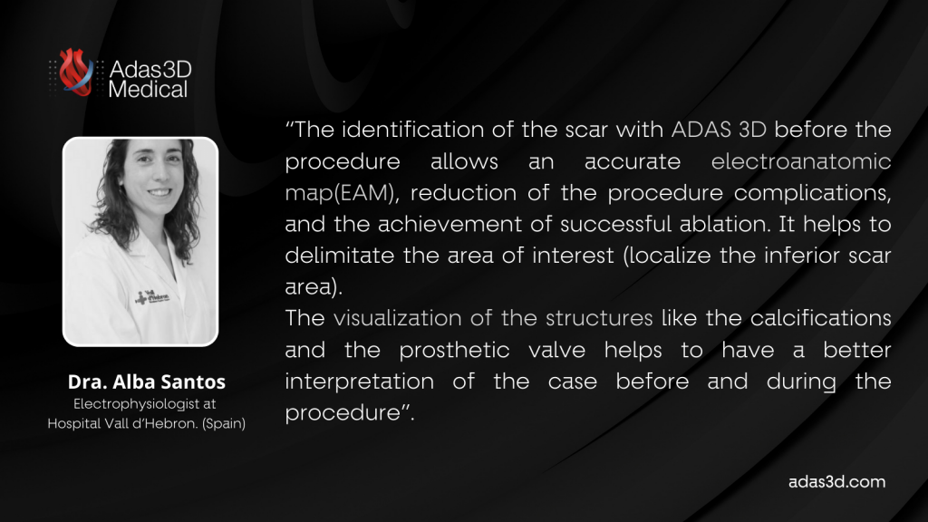
Thank you, Dra. Alba Santos, Gabriel Pascual, and all the Vall d’Hebron arrhythmia unit team for sharing this interesting case with us.
If you want to share your successful cases analyzed with ADAS 3D, please fill in this form.